Diseases of External Ear
As we have learned the anatomy of external ear in the previous post ,now we will check important high yield facts about external ear diseases.we divide the topic into:
- congenital disorders
- traumatic
- inflammatory
- neoplastic.
auricle/pinna:
bat ear(lop ear) :"lop"-to hang down loosely
- Definition: The external ear stands away from the head at a greater angle (Normal angle of the auricle to the median plane averages 25 degrees in boys and 18 degrees in girls). Lop ears are usually larger than normal ears.
- concha is large
- poorly developed antihelix & scapha.both these feature clearly seen in this picture.
- Associates anomalies: Commonly associated with the following:
- Ehlers-Danlos syndrome: Unusual facies, lop ears, hyperextensible joints, hip dislocation, inguinal hernia with autosomal recessive inheritance.
- Towns-Brocks syndrome: Lop ears, imperforate anus, hypoplastic kidney, ventricular septal defect, limb anomalies with autosomal dominant inheritance.
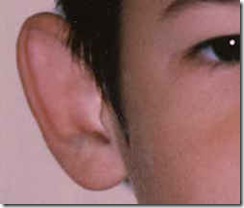
- treatment - by surgical correction -Bat ear correction or otoplasty, is a plastic surgery procedure designed to pull the ears closer to the head. Otoplasty involves reshaping, remodeling, and reforming the outer ear.
- complete absence of pinna
- part of "First arch syndrome"
- Definition: Macrotia means large ears.
- The auricle is usually very large but well shaped without other ear malformations.
- The most exaggerated portion is the scaphoid fossa.
- Microtia means small ears
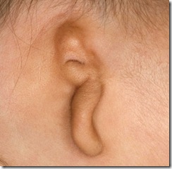
- associated anomalies :
| DIAGNOSIS | MINIMAL CRITERIA |
| microtia | Isolated microtia |
| Hemifacial microsomia | Unilateral microtia |
| Goldenhar syndrome | Unilateral microtia, small malformed mandible |
| Oculo-auriculo-vertebral dysplasia | Unilateral microtia, small malformed mandible, epibulbar dermoids, anomalies of the cervical spine |
- microtia-anotia is an essential component of isoretinoin embryopathy,
- important manifestation of thalidomide embryopathy, and
- can be part of the fetal alcohol syndrome and
- maternal diabetes embryopathy.
- Microtia-anotia occurs with a number of single gene disorders, such as Treacher-Collins syndrome, or chromosomal syndromes, such as trisomy 18.
- Rubella and other intrauterine infections
- Trisomy 21: Mean ear length and measured: expected ear length ratios are significantly lower in 75% of fetuses with trisomy 21
- Retinoic acid embryopathy
preauricular appendages:
- Definition: tags of skin with or without a cartilaginous base frequently located in the line drawn from tragus to angle of mouth.
- are often associated with microtia, melotia( Definition: Ear located on the cheek.) or oblique facial features
- due to a failure in the fusion of the primitive ear hillocks, or to a defective closure of the first branchial cleft
- characteristically located in or just in front of the anterior crus of the helix.
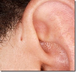
- The pits often discharge a cheesy substance, which consists of desquamated keratin debris.
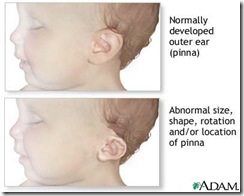
Associated anomalies: Commonly associated with the following
- Noonan syndrome: webbing of the neck, pectus excavatum, cryptorchidism, pulmonary stenosis. Ears are low-set with or without abnormal auricles. 25% have mental retardation. Koretzy et al. described an unusual type of pulmonary valvular dysplasia which showed a familial tendency with either affected parent and offspring or affected sibs. Other features were retarded growth, abnormal facies (triangular face, hypertelorism, low-set ears and ptosis of the eyelids)
- Pena Shokeir phenotype: Neurogenic arthrogryposis, pulmonary hypoplasia, hypertelorism with low-set malformed ears. 30% are stillborn. Majority of those liveborn die within the first month of life[15].
- Trisomy 18:Clenched hand, short sternum, low arch dermal ridge patterning on fingertips and low-set malformed auricles. Only 5% to 10% survive the first year as severely mentally defective individuals.
- Definition: In Agnathia-Synotia-Microstomia, the ears are very close to each other due to absence or hypoplasia of the mandible.
- The external ears assume a horizontal position with the lobules located near the midline.
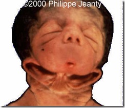
- "Bow-tie" ear due to failure of migration in Agnathia-Synotia-Microstomia
atresia:
| when occurs alone | when in association with microtia |
| due to failure of canalisation of ectodermal corethat fills the dorsal part of first branchial cleft | associated with abnormailities of middle ear ,inner ear |
| outer meatus-filled with fibrous tissue or bone | total atresia |
| deep meatus-normal |
collaural fistula:
- has two openings, the lower one situated in the neck between the angle of the mandible and the sternomastoid muscle, the upper in the floor of the external auditory canal or intertragal notch.
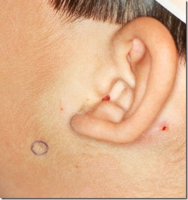
The lower opening of the collaural fistula is outlined with a blue circle .
traumatic :
auricle:
hematoma of auricle :
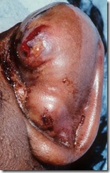

- collection of blood b/w auricular cartilage & perichondrium.
- this blood may clot & organise into "cauliflower ear"
- treatment -
- aspiration of hematoma & pressure dressing.
- if aspiration fails -incision & drainage
- prophylactic antibiotics.
laceration:
- cut in the pinna
- treatment: as fast as possible by stiching
- perichondrium - absorbable suture use
- skin - non-absorbable suture
- broad spectrum antibiotics
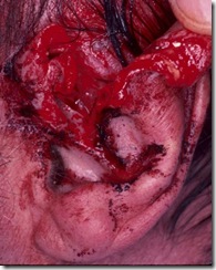
- The blood supply to the auricle is excellent and its metabolic demands are relatively low; therefore, primary reattachment is frequently successful, even with a tenuous pedicle.
- treatment :Management of these injuries consists of an early meticulous reimplantation, with minimal debridement and the use of low-dose heparin sodium to prevent thrombosis, appropriate antibiotic coverage to prevent infection, and multiple stab incisions to relieve venous congestion while revascularization is taking place.
- excessive scar at site of piercing
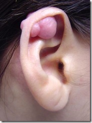
- usual sites-lobule or helix
{note-Hypertrophic scars remain within the confines of the wound and flatten spontaneously over one or more years. By contrast, keloids not only persist but frequently extend beyond the site of the original injury. Keloids occur more commonly in blacks than in whites and develop more frequently in the second and third decades of life.}
- treatment:
- surgical excision causes recurrence.
- to prevent recurrence-
- pre & post-operative radiation with total dose of 600-800 rad delivered in 4 divided doses
- local injection of steroid after excision.
| minor lacerations | major lacerations |
| from Q-tip injury | gun shot wounds,accidents |
| heal without sequelae | stenosis of ear canal is common problem |
ear drum:traumatic rupture
- trauma due to hair pin ,match stick
- sudden change in pressure- slap , forceful valsava procedure
- fracture of temporal bone
if it doesn't heal-tympanoplasty.
inflammatory disorders:
pinna:
| PERICHONDRITIS | RELAPSING POLYCHONDRITIS |
| infection secondary to laceration ,hematoma ,surgical incision or extension of infection from ear canal | autoimmune in origin |
| pseudomonas is important cause.others-Staphylococcus aureus, and Streptococcus pyogenes | does not involve any bacteria |
| symptoms-red ,hot ,painful pinna & stiff | same & other cartilages like tracheal ,nasal septum ,coastal cartilages also involved. |
| later abscess may form | no |
| treatment-early stage:systemic antibiotics like fluoroquinolones-ciprofloxacin are drug of choice | Treatment is with oral corticosteroids. |
| if an abscess is present, surgical incision and drainage often are necessary. |
{note:in both these conditions lobule is unaffected thus differentiating from cellulitis.}
chondrodermatitis nodularis chronica helicis:
- small benign painful nodules on free border of helix

- most authorities believe it is caused by prolonged and excessive pressure.check this on emedicine
- treatment:surgical excision of underlying cartilage.


I like your article and it really gives an outstanding idea that is very helpful for all the people on web.
ReplyDeleteHearing Aid